How Portable Dental X-Ray Machines Work
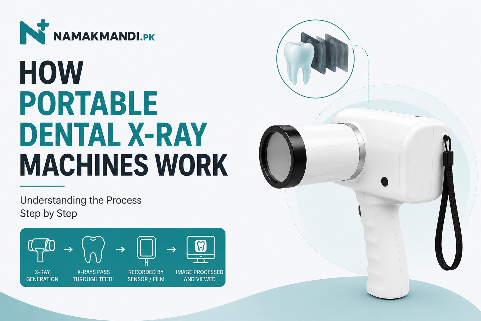
Portable dental X-ray machines provide dentists with a compact way to produce intraoral dental radiographs without relying on a permanently wall-mounted X-ray generator. Despite their smaller size and handheld design, the basic science behind them is similar to that used in conventional dental X-ray equipment.
The portable machine itself does not usually store or create the final visible image. Its main job is to generate a controlled X-ray beam. A separate image receptor—such as a digital sensor, PSP plate, or dental X-ray film—receives the X-rays after they pass through the patient’s teeth and surrounding tissues.
Understanding this process can help dentists, dental students, clinic owners, and equipment buyers understand what the machine does, what additional equipment may be required, and how different dental imaging setups work.
What Is a Portable Dental X-Ray Machine?
A portable dental X-ray machine is a compact X-ray generator designed primarily for intraoral dental radiography.
Unlike a traditional unit mounted permanently to a wall or supporting structure, a portable unit can be moved between treatment areas. Many modern models are designed to be held by a trained operator during imaging and use a rechargeable battery as their power source.
Portable units may be used to produce common intraoral radiographs such as periapical and bitewing images, depending on the clinical indication, equipment, receptor, and technique.
The important point is:
X-ray machine = generates the X-rays
Sensor, PSP plate, or film = records the radiographic information
They are separate components of the imaging process.
Main Parts of a Portable Dental X-Ray Machine
The exact design differs between manufacturers, but a typical handheld dental X-ray machine contains several important components.
1. X-Ray Tube
Inside the machine is an X-ray tube where electrical energy is converted into X-rays.
The tube itself is contained within protective housing, so the operator does not interact directly with it.
2. High-Voltage Generator
Producing X-rays requires a substantial electrical potential within the tube. The generator provides the electrical conditions required for X-ray production.
Modern portable units package this technology into a relatively small housing.
3. Rechargeable Battery
Many handheld dental X-ray machines use rechargeable lithium-based batteries.
The battery supplies the electrical energy required for operation, allowing the unit to be used without remaining connected to a wall outlet during every exposure.
Battery capacity, charging time and the number of exposures achievable per charge can differ considerably between models.
4. Position-Indicating Device
The cylindrical or cone-shaped section at the front of the machine helps direct the X-ray beam toward the intended area.
Correct alignment of the X-ray source, teeth and image receptor is important for obtaining a useful radiograph.
5. Exposure Controls
Portable machines normally provide controls that allow the operator to select appropriate exposure parameters.
Depending on the model, settings may be selected manually or through predefined programs based on factors such as the tooth region or receptor being used.
6. Backscatter Shield
Many handheld dental X-ray units incorporate a protective shield around the front portion of the device.
Its purpose is to help reduce the amount of scattered radiation reaching the operator during properly performed exposures.
The design and correct positioning of this shield are important considerations when evaluating handheld equipment.
How Does a Portable Dental X-Ray Machine Work?
The complete imaging process can be understood in a series of steps.
Step 1: The Image Receptor Is Positioned
Before taking the radiograph, an appropriate image receptor is positioned relative to the teeth being examined.
This may be:
- An intraoral digital sensor
- A PSP plate
- Conventional dental X-ray film
Positioning devices may be used to help maintain appropriate alignment.
Step 2: The X-Ray Machine Is Positioned
The portable X-ray generator is aligned with the tooth or region of interest and the receptor.
Correct positioning matters because poor horizontal or vertical angulation can distort the resulting image or cause important structures to be missed.
With a handheld system, maintaining stable positioning during exposure is particularly important.
Step 3: Exposure Parameters Are Selected
The machine needs to deliver an appropriate amount and quality of X-radiation for the examination.
Depending on the unit, the operator may select or adjust parameters such as:
- Tube voltage (kV)
- Tube current (mA)
- Exposure time
Some machines simplify this process through preset exposure programs.
The appropriate settings depend on the equipment, receptor, anatomical region and manufacturer recommendations.
Step 4: X-Rays Are Generated
When the exposure is activated, electrical energy is applied inside the X-ray tube.
Electrons are accelerated toward a metal target. When these high-speed electrons interact with the target, part of their energy is converted into X-ray photons.
Most of the energy involved is converted into heat rather than X-rays, which is why X-ray equipment must be designed to manage heat safely.
Step 5: The X-Ray Beam Passes Through the Teeth
The useful X-ray beam leaves the machine and travels through the area being examined.
Different tissues and materials absorb different amounts of X-radiation.
Dense structures absorb more of the beam, while less-dense structures allow more X-rays to pass through.
Step 6: The Receptor Records the Information
After passing through the teeth and surrounding structures, the remaining X-rays reach the image receptor.
The pattern of X-rays reaching that receptor contains the information needed to create the radiographic image.
What happens next depends on whether the clinic is using a digital sensor, PSP plate or conventional film.
Why Do Teeth and Bone Look Different on an X-Ray?
A dental radiograph is essentially based on differences in X-ray attenuation.
Dense materials absorb more X-rays and therefore generally appear lighter or more radiopaque on the resulting image.
Less-dense areas allow a greater proportion of X-rays to reach the receptor and tend to appear darker or more radiolucent.
For example:
| Structure/Material | Typical Radiographic Appearance |
|---|---|
| Enamel | Very radiopaque/light |
| Dentin | Less radiopaque than enamel |
| Bone | Varies according to density and anatomy |
| Metal restorations | Often highly radiopaque |
| Air spaces | Dark/radiolucent |
| Soft tissues | Relatively low contrast |
Dentists interpret these patterns alongside clinical findings to assess teeth and surrounding structures.
How Portable Dental X-Ray Machines Work With Digital Sensors
A digital intraoral sensor can be positioned inside the patient’s mouth in much the same location where conventional dental film would otherwise be placed.
The portable X-ray machine is positioned and the exposure is made.
The process is essentially:
Portable X-ray generator → Patient’s teeth → Digital sensor → Imaging system → Displayed radiograph
The sensor converts the received X-ray information into an electronic signal. The associated computer and imaging software process that information into an image that can be viewed on a screen.
One major advantage of direct digital sensors is speed. There is no conventional chemical film-development stage.
Does the Portable X-Ray Machine Include the Sensor?
Not necessarily.
This is an important consideration when purchasing dental imaging equipment.
A seller may advertise a portable dental X-ray machine, but the quoted price may refer only to the X-ray generator.
The digital sensor, imaging software, computer equipment and accessories may be separate purchases.
Buyers should therefore check exactly what is included in a package rather than assuming that a portable X-ray machine automatically provides a complete digital radiography system.
How Portable Dental X-Ray Machines Work With Dental X-Ray Film
Portable X-ray generators can also be used with conventional intraoral dental X-ray film when the particular equipment and exposure settings are suitable.
The film is positioned inside the patient’s mouth and exposed to X-rays after the beam passes through the teeth.
Unlike a direct digital sensor, conventional film does not immediately display an image.
It must be appropriately processed before the radiograph becomes visible.
The workflow is therefore:
Portable X-ray generator → Teeth → Dental X-ray film → Film processing → Radiograph
Film-based imaging may remain relevant in clinics that have not transitioned completely to digital radiography or where conventional film continues to form part of the existing workflow.
What About PSP Plates?
PSP, or photostimulable phosphor plates, provide another option.
They resemble conventional film in terms of being relatively thin receptors, but the exposed plate is subsequently placed into a dedicated scanner.
The scanner reads the stored radiographic information and converts it into a digital image.
The workflow becomes:
X-ray machine → PSP plate → PSP scanner → Computer → Digital image
This makes PSP an intermediate type of workflow between immediate direct digital sensors and traditional film processing.
What Controls the X-Ray Exposure?
Three concepts commonly encountered in dental radiography are kV, mA and exposure time.
Kilovoltage (kV)
Kilovoltage affects the energy and penetrating ability of the X-ray beam.
Increasing kV generally produces X-rays with greater penetrating power.
Milliamperage (mA)
Tube current relates to the number of electrons flowing through the X-ray tube and therefore influences the quantity of X-rays produced.
Exposure Time
Exposure time determines how long X-rays are produced during an exposure.
Portable dental X-ray machines may simplify these parameters through presets, but the underlying principles remain relevant.
The operator should follow the manufacturer’s recommended exposure protocols and applicable professional guidance rather than adjusting settings arbitrarily.
What Happens With the X-Rays That Don’t Reach the Sensor?
Not every X-ray photon travels directly through the patient and reaches the receptor.
Some are absorbed, while others can change direction after interacting with matter. This latter phenomenon contributes to scattered radiation.
For handheld dental X-ray equipment, managing scatter is particularly relevant because the operator may remain near the unit during exposure.
This is why equipment design and correct operating technique matter.
How Do Portable X-Ray Machines Protect the Operator?
Radiation protection involves more than one feature.
A well-designed handheld system may incorporate:
Protective Housing
The X-ray tube and associated components are enclosed within housing designed to limit radiation leakage outside the intended beam.
Beam Limitation
The useful X-ray beam is directed toward a defined area rather than allowing radiation to travel unnecessarily in every direction.
Backscatter Shield
A properly designed and positioned backscatter shield helps reduce scattered radiation reaching the operator.
Short, Controlled Exposures
The equipment produces a controlled exposure according to selected parameters.
Correct Operating Technique
Equipment features alone are not enough. Appropriate training, positioning, maintenance, quality assurance and adherence to applicable radiation-protection requirements remain important.
What Happens After the X-Ray Is Taken?
This depends entirely on the receptor.
With a Direct Digital Sensor
The image can generally be displayed through the connected imaging system shortly after exposure.
With a PSP Plate
The exposed plate must be placed into a compatible scanner before the image can be displayed digitally.
With Conventional Film
The film requires appropriate processing before the radiograph can be viewed.
Therefore, buying a portable X-ray generator alone does not determine whether a clinic has a digital or conventional radiography workflow. The receptor system plays a major role.
Does a Portable X-Ray Machine Work Differently From a Wall-Mounted Machine?
At the fundamental level, both produce X-rays using the same basic physical principles.
Both require:
- An X-ray source
- Electrical energy
- Controlled X-ray generation
- Beam direction
- An image receptor
The biggest differences relate to the physical design, power source, positioning and clinical workflow.
A traditional intraoral unit normally remains attached to a wall or supporting structure. The tube head is moved into position around the patient.
With a handheld portable unit, the generator itself can be carried and positioned by the operator.
The underlying goal remains the same: deliver an appropriately controlled X-ray exposure through the region of interest to an image receptor.
What Can Portable Dental X-Rays Be Used For?
Intraoral dental radiographs can support the assessment of many dental conditions and procedures.
Depending on clinical need and the type of radiograph, imaging may assist with:
- Examining individual teeth
- Assessing tooth roots
- Evaluating surrounding bone
- Identifying certain forms of dental decay
- Assessing periodontal bone levels
- Root canal procedures
- Evaluating impacted teeth
- Checking selected dental restorations
- Treatment planning
- Follow-up assessment
The decision to take a radiograph should be based on clinical need rather than taking X-rays routinely without justification.
Portable X-Ray Machine vs Digital Sensor
Because these two products are often mentioned together, the distinction is worth making clear.
| Portable X-Ray Machine | Digital Dental Sensor |
|---|---|
| Generates X-rays | Detects X-rays |
| Positioned outside the mouth | Usually positioned inside the mouth |
| Contains an X-ray tube | Contains an imaging detector |
| Creates the exposure | Records radiographic information |
| Can potentially work with different receptors | Requires an X-ray source |
| Does not itself replace the receptor | Does not generate X-rays |
A dentist establishing a digital imaging setup generally needs both an X-ray generator and an appropriate digital receptor system.
A Complete Portable Digital Dental X-Ray Setup
A basic digital setup may therefore involve:
1. Portable dental X-ray machine
Generates the X-ray beam.
2. Intraoral digital sensor
Captures the radiographic information.
3. Computer or compatible device
Receives and processes the data.
4. Imaging software
Displays and manages the resulting radiographs.
Depending on the equipment, additional accessories and positioning devices may also be required.
Understanding these components is particularly useful when comparing supplier quotations because a low advertised price may cover only one part of the complete system.
Frequently Asked Questions
Does a portable dental X-ray machine take digital X-rays?
The generator produces X-rays rather than determining whether the final image is digital. When used with a compatible digital sensor or PSP system, it can form part of a digital dental radiography setup.
Does a portable X-ray machine need a sensor?
It needs an appropriate image receptor. That receptor could be a digital sensor, PSP plate or suitable dental X-ray film, depending on the equipment and intended workflow.
How quickly can a portable X-ray produce an image?
With a direct digital sensor, the image can generally become available very quickly through the associated imaging system. PSP plates require scanning, while conventional film requires processing.
Can portable dental X-ray machines use normal dental X-ray film?
Some portable units can be used with conventional intraoral dental X-ray film when appropriate exposure settings and procedures are used. The specifications of the particular machine should be checked.
Are portable and handheld dental X-ray machines the same?
The terms are sometimes used interchangeably, but not every portable system necessarily has to be held in the operator’s hands during exposure. Product design should therefore be checked rather than relying solely on the term “portable.”
Does a portable X-ray machine need electricity?
Many handheld models use rechargeable batteries, allowing them to operate without a mains connection during each exposure. They still require an appropriate power source for charging.
Is the X-ray technology different from a traditional dental X-ray?
The basic principle of X-ray generation is similar. The major differences concern the machine’s size, mounting, power supply, positioning and workflow.
Final Thoughts
Portable dental X-ray machines take a technology traditionally associated with fixed installations and package it into a more compact and mobile system.
The process begins inside the X-ray tube, where electrical energy is used to generate X-rays. The beam passes through the patient’s teeth and surrounding tissues before reaching a digital sensor, PSP plate or dental X-ray film. Differences in how those structures absorb X-rays create the information used to form the radiographic image.
What makes a portable system different is therefore not an entirely different type of X-ray physics, but the way the generator is designed, powered and positioned.
For dentists considering a portable unit, understanding this distinction is useful when comparing equipment. The X-ray generator, digital sensor and imaging software may all be separate components, and compatibility between them should be considered before purchasing a complete dental radiography setup.